Date:16/07/18
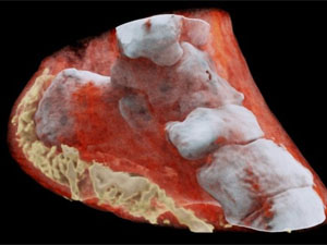 Medical X-ray scans have long been stuck in the black-and-white, silent-movie era. Sure, the contrast helps doctors spot breaks and fractures in bones, but more detail could help pinpoint other problems. Now, a company from New Zealand has developed a bioimaging scanner that can produce full color, three dimensional images of bones, lipids, and soft tissue, thanks to a sensor chip developed at CERN for use in the Large Hadron Collider.
Mars Bioimaging, the company behind the new scanner, describes the leap as similar to that of black-and-white to color photography. In traditional CT scans, X-rays are beamed through tissue and their intensity is measured on the other side. Since denser materials like bone attenuate (weaken the energy) of X-rays more than soft tissue does, their shape becomes clear as a flat, monochrome image.
But for the new technology, which Mars calls "Spectral CT," the sensor can measure the attenuation of specific wavelengths of the X-rays as they pass through different materials. After running the spectroscopic data through specific algorithms, a 3D color image is generated that clearly shows muscle, bone, water, fat, disease markers – and even a watch. The end results are unnerving, like someone's sculpted a detailed clay model of your insides.
At the heart of the Spectral CT scanner is a Medipix3 chip. This device, which detects and counts every individual particle that hits each pixel on the sensor, was originally developed at CERN to precisely track particles in the Large Hadron Collider.
A small version of the device has been tested to see how well it can diagnose bone and joint health, spot cancer, and pick up early markers for vascular diseases. So far, the results have been promising, the team says.
"In all of these studies, promising early results suggest that when spectral imaging is routinely used in clinics it will enable more accurate diagnosis and personalization of treatment," says Anthony Butler, one of the creators of the 3D scanner.
Clinical trials are set to begin over the next few months in New Zealand, as the Mars scanner is put to work on orthopaedic and rheumatology patients.


CERN chip enables first 3D color X-ray images of the human body
Medical X-ray scans have long been stuck in the black-and-white, silent-movie era. Sure, the contrast helps doctors spot breaks and fractures in bones, but more detail could help pinpoint other problems. Now, a company from New Zealand has developed a bioimaging scanner that can produce full color, three dimensional images of bones, lipids, and soft tissue, thanks to a sensor chip developed at CERN for use in the Large Hadron Collider.Mars Bioimaging, the company behind the new scanner, describes the leap as similar to that of black-and-white to color photography. In traditional CT scans, X-rays are beamed through tissue and their intensity is measured on the other side. Since denser materials like bone attenuate (weaken the energy) of X-rays more than soft tissue does, their shape becomes clear as a flat, monochrome image.
But for the new technology, which Mars calls "Spectral CT," the sensor can measure the attenuation of specific wavelengths of the X-rays as they pass through different materials. After running the spectroscopic data through specific algorithms, a 3D color image is generated that clearly shows muscle, bone, water, fat, disease markers – and even a watch. The end results are unnerving, like someone's sculpted a detailed clay model of your insides.
At the heart of the Spectral CT scanner is a Medipix3 chip. This device, which detects and counts every individual particle that hits each pixel on the sensor, was originally developed at CERN to precisely track particles in the Large Hadron Collider.
A small version of the device has been tested to see how well it can diagnose bone and joint health, spot cancer, and pick up early markers for vascular diseases. So far, the results have been promising, the team says.
"In all of these studies, promising early results suggest that when spectral imaging is routinely used in clinics it will enable more accurate diagnosis and personalization of treatment," says Anthony Butler, one of the creators of the 3D scanner.
Clinical trials are set to begin over the next few months in New Zealand, as the Mars scanner is put to work on orthopaedic and rheumatology patients.
Views: 425
©ictnews.az. All rights reserved.
Similar news
- The mobile sector continues its lead
- Facebook counted 600 million active users
- Cell phone testing laboratory is planned to be built in Azerbaijan
- Tablets and riders outfitted quickly with 3G/4G modems
- The number of digital TV channels will double to 24 units
- Tax proposal in China gets massive online feedback
- Malaysia to implement biometric system at all entry points
- Korea to build Green Technology Centre
- Cisco Poised to Help China Keep an Eye on Its Citizens
- 3G speed in Azerbaijan is higher than in UK
- Government of Canada Announces Investment in Green Innovation for Canada
- Electric cars in Azerbaijan
- Dominican Republic Govt Issues Cashless Benefits
- Spain raises €1.65bn from spectrum auction
- Camden Council boosts mobile security